- info@vishnunetralaya.com
- 404, akashdeep plaza, 4th floor, Golmuri, Jamshedpur - 831003
- +91 9835316814
Retinal Diseses
Retinal Diseses
Retinal diseases vary widely, but most of them cause visual symptoms. Retinal diseases can affect any part of your retina, a thin layer of tissue on the inside back wall of the eye.
The retina contains millions of light-sensitive cells, called rods and cones, and other nerve cells that receive and organize visual information. The retina sends this information to the brain through the optic nerve, enabling you to see.
Treatment is available for some retinal diseases. Depending on your condition, treatment goals may be to stop or slow the disease. This may help preserve, improve or restore your vision. Untreated, some retinal diseases can cause severe vision loss or blindness.
Types
Common retinal diseases and conditions include:
Retinal tear: A retinal tear occurs when the clear, gel-like substance in the center of your eye, called vitreous, shrinks and tugs on the thin layer of tissue lining the back of your eye, called the retina. This can cause a tear in the retinal tissue. It's often accompanied by the sudden onset of symptoms such as floaters and flashing lights.
Retinal detachment: A retinal detachment is defined by the presence of fluid under the retina. This usually occurs when fluid passes through a retinal tear, causing the retina to lift away from the underlying tissue layers.
Diabetic retinopathy: If you have diabetes, the tiny blood vessels in the back of your eye can deteriorate and leak fluid into and under the retina. This causes the retina to swell, which may blur or distort your vision. Or you may develop new, irregular capillaries that break and bleed. This also worsens your vision.
Epiretinal membrane: Epiretinal membrane is a delicate tissue-like scar or membrane that looks like crinkled cellophane lying on top of the retina. This membrane pulls up on the retina, which distorts your vision. Objects may appear blurred or crooked.
Macular hole: A macular hole is a small defect in the center of the retina at the back of the eye, called the macula. The hole may develop from atypical traction between the retina and the vitreous, or it may follow an injury to the eye.
Macular degeneration: In macular degeneration, the center of the retina begins to deteriorate. This causes symptoms such as blurred central vision or a blind spot in the center of the visual field. There are two types — wet macular degeneration and dry macular degeneration. Many people will first have the dry form, which can progress to the wet form in one or both eyes.
Retinitis pigmentosa: Retinitis pigmentosa is an inherited degenerative disease. It slowly affects the retina and causes loss of night and side vision.

- Vishnu Netralaya has the unique distinction of starting the First Speciality Retina unit at Jamshedpur. It was founded by a Retina consultant trained at Sankara Nethralaya ,Chennai and at RIO, Patna.
- As a part of our protocol, we do a thorough Retina Check up of every OPD patients via Indirect Ophthalmoscopy

- Zeiss CIRRUS® OCT 6000: It Performs 1,00,000 scans per second with HD imaging detail and a wide field of view providing detailed information of all 10 layers of Retina at a cellular level.
- Zeiss CIRRUS® OCT Angiography : A non-invasive procedure to determine the Vascular integrity of Retina and Choroid. Since no dye is injected, it eliminates the dye related complications and is very comfortable to the patient.
- Topcon Fundus Fluorescein Angiography (FFA) an absolutely essential modality to locate leaking Blood vessels in retina
- Fundus Photography (TOPCON) : To record the extent of Retinal Pathology and monitor its resolution or progress

- Laser Photocoagulation: Pioneered in Jamshedpur by Vishnu Netralaya. We use the most advanced Green LASER (Double Frequency) module that causes least discomfort to the patients. We have the Facility of both
- - Slit Lamp LASER for ambulatory patients who can sit easily, and
- - LASER Indirect Ophthalmoscope for very sick, elderly and infant patient
- Fundus Photography (TOPCON): To record the extent of Retinal Pathology and monitor its resolution or progress

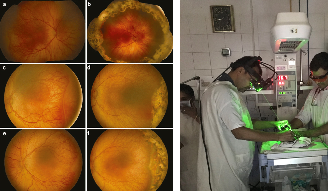
- We are the Pioneering and Leading Centre in Jamshedpur in screening ,diagnosing and Treating Retinopathy of Prematurity (ROP) in Preterm Babies
- LASER Procedure of a Preterm Baby in Neonatal ICU
- Before and after laser photocoagulation for retinopathy of prematurity (ROP)
- It is an testimony to our success and leadership in this field that the first institute that patients requiring Retina Treatment, in Jamshedpur, think of is Vishnu Netralaya
